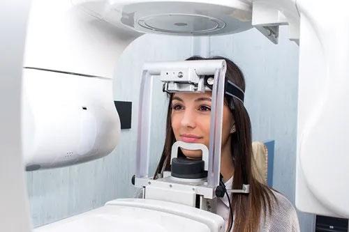
3D Cone Beam Scan
3D cone beam scans are a type of x-ray that can help dentists investigate the structure of your mouth in situations where traditional two-dimensional xrays are not sufficient. This scanning method produces high-fidelity images of your teeth and gums, allowing dental professionals to explore them in three-dimensions for better diagnosis and treatment.
How Does 3D Cone Beam Scanning Work?
 3D cone beam scanning is so-named because of the shape of the x-ray emissions as they emerge from the scanning device. Traditional equipment scans used a fan-shaped beam but cone beam scanning, as the name implies, emits a funnel-shaped beam.
Cone beam CT scanners are square-shaped machines that rotate around patients’ heads, either while they stand up or sit down. Usually, a rotating C-arm containing the x-ray source circles the head, scanning it from all angles, while detectors collect information.
As the x-ray source moves around the patients’ head, it produces a large number of high-quality images, showing details of both hard and soft tissues at varying depths. Computer software then stitches all these together to produce a navigable three-dimensional model that dentists can then use to develop treatment plans.
Originally, researchers developed dental cone technology to make digital x-ray machines small enough to fit in dental offices. Devices that used the technology were less expensive than older models using classical xray scanning technology. Furthermore, they offered higher fidelity images of facial structures, including sinus and nasal cavities and the jaw but with lower radiation exposure compared to conventional CT.
Preparation for 3D cone beam scanning is minimal. Dentists request that patients remove anything that could interfere with the procedure, including metal jewelry, eyeglasses, hearing aids and hairpins. In some cases, they may also remove removable prosthetics from your mouth, such as dentures.
The procedure typically lasts between 20 and 40 seconds, depending on the equipment and area being scanned. Regional scans — those that focus on a specific area of the mandible or maxilla — typically take around 10 seconds.
Why Do Dentists Use 3D Cone Beam Scans?
In some cases, 2D x-rays may not provide dentists with sufficient information to proceed with an optimal treatment plan. In these situations, dentists may call on 3D beam scanning. 3D cone beam scanning tends to be more useful in complex oral health conditions. For instance, dentists will sometimes use it when planning impacted teeth surgery. Cone beam scanning provides more detailed information about the orientation of the impacted tooth and surrounding soft tissue, such as nerves, that are at risk of damage during surgery.
Other applications include:
- Accurately placing implant posts into the jawbone during implant surgery
- Planning for reconstructive surgery of either the jaw or dentition
- Diagnosis of jaw conditions, such as temporomandibular joint disorder (TMJ)
- Detailed evaluation of perioral cavities, such as sinuses
- Identification of the source of jaw pain
- Identification of oral or jaw tumors
What Are The Benefits Of 3D Cone Beam Scans?
3D cone beam mouth scans offer various benefits over conventional xrays and CT scans, many of which we have hinted at above.
Higher-Fidelity Scans
Dentists are able to offer patients superior treatment when they have a better understanding of the structure of their face, jaw and dentition. 3D cone beam scans make it easier to explore three-dimensional images of a patient’s mouth than 2D images.
Capital Oral & Facial uses state-of-the-art, in-house digital imaging 3D cone beam technology. It allows our dentists to create an accurate picture of the interior of your head. Proper scanning lets us map out the location of sensory nerves, select the right implant length for your jawbone, improve placement of oral prosthetics, and avoid penetration of implants into the sinus. It also improves our ability to identify the origin of pathology and pain, including potential causes of sleep apnea.
Reduced Radiation
Dental cone beam CT scans produce images of a similar quality to conventional CT. However, because of the cone-shaped nature of their beams, they expose patients to considerably less scatter radiation.
Evidence suggests that cone beam scanners expose patients to between 18 and 200 µSv — the equivalent of between 2.2 and 24.4 days of annual per capita background radiation. By contrast, a conventional CT scanner might expose patients to 860 µSv, which equates to more than 105 days of background radiation. This means that the radiation dose from cone beam scanners is roughly comparable to what humans are exposed to simply living on Earth.
Once the procedure is complete, no radiation remains in the patient’s body. There are no known immediate side-effects from the procedure.
Quick And Easy
3D cone beam scanning is also painless, quick and easy. Scanning only takes a few seconds and, once the process is over, dentists have all the details they need about the patient to proceed with the patient.
Fast Results
Once dentists have the output from the 3D cone scanning machine, they can immediately recommend a course of treatment. In some instances, you may be able to undergo a procedure immediately.
What Are The Risks?
While 3D cone beam scanning is one of the best teeth xray options out there, there are some risks.
- Pregnant women should not use 3D cone beam scanners
- 3D cone beam scanning still exposes patients to a small quantity of radiation. However, the intensity of the radiation is many times less than a CT scan
- Children are more sensitive to 3D cone beam scanning so dentists will only use it if absolutely necessary and at a low dose
Get 3D Cone Beam Scanning
Capital Oral & Facial offers 3D Cone Beam scanning (CBCT) to support various treatments and improve care. Our state-of-the-art equipment gets to work immediately, with pain-free scans lasting just a few seconds. Become a valued patient at our clinic today and experience the benefits for yourself.

